Home >> Foot & Ankle Services
Foot and Ankle Services
The ankle joint is a synovial joint, of the lower limb, that is formed by 3 bones:
the tibia and fibula of the leg, and the talus of the foot:
The tibia and fibula are bound together by strong tibio-fibular ligaments in the front and back. Together, they form a bracket shaped socket, covered in hyaline cartilage. This socket is known as a mortise. The body of the talus fits snugly into the mortise formed by the bones of the leg. The articulating part of the talus is wedge shaped, being broad anteriorly (in front) & narrow posteriorly (at back).

Functionally, ankle is a hinge joint, that permits movements to occur in one plane only. These ankle movements are dorsiflexion and plantarflexion
Dorsiflexion is when the foot and toes point downwards. During dorsiflexion, the anterior part of the talus is held in the mortise, and the joint is more stable. It is caused by the actions of muscles of the anterior compartment of the leg (tibialis anterior, extensor hallucis longus and extensor digitorum longus).
Plantarflexion is when the foot and toes point upwards (ankle and toes point upwards) movements. During plantarflexion, the posterior part of the talus is held in the mortise, and the joint is less stable. It is caused by the actions of muscles of the posterior compartment of the leg (gastrocnemius, soleus, plantaris and posterior tibialis).
There are two main sets of ligaments: the medial (inner) and lateral (outer) ligaments.
The medial ligament (or deltoid ligament) is attached to the medial malleolus – a bony prominence projecting from the medial aspect of the distal tibia.
It consists of four ligaments, which fan out from the malleolus, attaching to the talus, calcaneus and navicular bones. The primary action of the medial ligament is to resist over-eversion (outward movement) of the foot.

The lateral ligament originates from the lateral malleolus (a bony prominence projecting from the lateral aspect of the distal fibula). It is comprised of three distinct and separate ligaments:
Anterior talofibular – spans between the lateral malleolus and lateral aspect of the talus.
Posterior talofibular – spans between the lateral malleolus and the posterior aspect of the talus.
Calcaneofibular – spans between the lateral malleolus and the calcaneus.
It resists over-inversion (inward movement) of the foot.
The feet are flexible structures of bones, joints, muscles, and soft tissues that let us stand upright and perform activities like walking, running, and jumping.
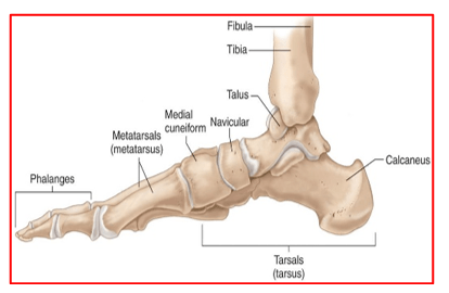
The foot is divided into three sections: Hind-foot, Mid-foot and Fore-foot.
The two bones that make up the back part of the foot (sometimes referred to as the hindfoot) are the talus and the calcaneus (heel bone). The talus is connected to the calcaneus at the subtalar joint that allows the foot to rock from side to side (inversion and eversion movements – foot turning inwards and outwards respectively)
The midfoot is a pyramid-like collection of 5 bones that work together as a group and form the arches of the feet. These include the three cuneiform bones, the cuboid bone, and the navicular bone. There are multiple joints, between the tarsal bones. When the foot is twisted in one direction by the muscles of the foot and leg, these bones lock together and form a very rigid structure. When they are twisted in the opposite direction, they become unlocked and allow the foot to conform to whatever surface the foot is contacting.
The tarsal bones are connected to the five long bones of the foot called the metatarsals. The two groups are fairly rigidly connected, without much movement at the joints.
The forefoot contains the five toes (phalanges) and the five longer bones (metatarsals). The joint between the metatarsals and the first phalanx of each toe is called the metatarso-phalangeal (MTP) joint. These joints form the ball of the foot, and movement in these joints is very important for a normal walking pattern. Not much motion occurs at the joints between the bones of the toes.
The big toe, or hallux, is the most important toe for walking, and the first MTP joint is a common area for problems in the foot.
Muscles, tendons, and ligaments run along the surfaces of the feet, allowing the complex movements needed for motion and balance.
Achilles tendon is the most important tendon for walking, running, and jumping. It attaches the calf muscles to the heel bone to allow us to rise up on our toes.
Tibialis posterior tendon attaches one of the smaller muscles of the calf to the underside of the foot. This tendon helps support the arch and allows us to turn the foot inward.
Tibialis anterior tendon allows us to raise the foot.
Two peroneal tendons (longus & brevis) run behind the outer bump of the ankle (lateral malleolus) and attach to the outside edge of the foot. These two tendons help turn the foot outward.
The toes have tendons attached on the bottom that bend the toes down (flexor tendons) and attached on the top of the toes that straighten the toes (extensor tendons.
Foot & Ankle disorders can result from damage to bones, muscles, or soft tissues.
Common foot & ankle disorders are:
- Sprains (injury to ligaments)
- Fractures
- Tendonitis (inflammation of the tendons) or tendon ruptures
- Arthritis (chronic inflammation of joints)
There are several causes for ankle disorders. This includes running, jumping, and overuse.
Common causes of ankle sprains and fractures include:
- twisting or rotating the ankle beyond the normal range of motion
- tripping or falling
- landing on the foot with increased force
Injuries that can lead to tendonitis in the ankle or Achilles tendonitis can be caused by:
- lack of conditioning for the muscles in the leg and foot
- excess strain on the Achilles tendon that connects the calf muscles to the heel
- bone spurs in the heel that rub on the Achilles tendon
- untreated flat feet leading to additional stress on the posterior tibial tendon
Different types of arthritis (inflammation of joints and tissues) can also affect the foot and ankle:
Osteoarthritis is a degenerative type of arthritis that typically begins in middle age and slowly progresses. Over time, cartilage between the ankle bones becomes worn down resulting in joint pain, stiffness and reduced mobility.
Rheumatoid arthritis is an autoimmune inflammatory disease that affects many joints in the body. The joint cartilage gets damaged due to the disease.
Post-traumatic arthritis occurs after an injury to the joint surfaces of the foot or ankle. Stress from the injury can cause the affected joints to become swollen, stiff, deformed and arthritic.
Gout can affect ankle or the big toe joint leading to repeated episodes of pain, swelling and inflammation of the joint, leading to arthritis of the joints
Common foot and ankle disorders seen by Orthopaedic surgeons:
- Ankle sprains and instability
- Ankle fractures
- Osteochondral injuries of the talus
- Ankle impingement
- Ankle arthritis
- Achilles tendinitis and ruptures
- Posterior tibial tendinitis & Flatfeet
- Calcaneal, Talus, Tarsal, Metatarsal and Phalangeal fractures
- Hind Foot, Mid foot and forefoot arthritis
- Cavus (high arched) foot
- Diabetic foot
- Gout
- Deformities of the big toes – hallux valgus, rigidus and varus
- Claw toes & hammer toes
- Plantar fasciitis
- Metatarsalgia
- Morton’s neuroma
- In-growing toenails
- Corn, calluses & warts
- Fungal infections of toes
Book An Appointment
Private Clinics : Locations & Directions
London Joints Clinic (Pune)
Address
Office S 5, 2nd Floor, North Block, Sacred World Mall,
Opp Sacred Heart Township, Near Jagtap Chowk,
Wanawadi, Pune 411040
Monday to Saturday
6 PM to 9 PM
Appointments
Hospitals OPDs : Locations & Directions
Jupiter Hospital (Baner)

Address
Lane 3, Baner- Balewadi Road,
Prathamesh Park,
Baner, Pune 411 045
Monday to Saturday 11 AM to 4 PM
Appointments
Contact us
Dr Anand Jadhav has a centralised appointment system for all locations across various hospitals and clinics in Pune & PCMC areas
Appointment Bookings & Requests can be made by any method :

