Flatfoot deformity is a condition characterized by low or absent foot arches. It makes the foot appear flat at the sole when standing and can cause foot pain and difficulty in walking.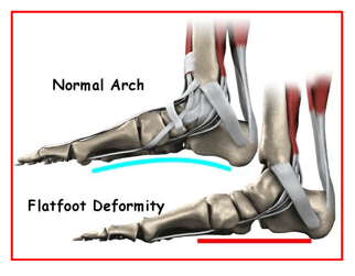
Flat feet are a common condition. It is normal in infants and toddlers.
Flat feet occur because the tissues holding the joints in the foot together (called capsules and tendons) are loose. These tissues gradually tighten and form an arch as children grow older. This will take place by the time the child is 2 or 3 years old. Most people have normal arches by the time they are adults.
In some people the arch may never form.
Some hereditary conditions patients have more elastic tissue in their body which causes tendons to be loose.
Ehlers-Danlos Syndrome
Marfan Syndrome
People born with these conditions have flat feet.
Aging, feet injuries, posterior tibial tendon dysfunction or illness like diabetes can cause flat feet to develop in a person who has already formed arches. This type of flat foot may occur only on one side.
Rarely, painful flat feet in children may be caused by a condition in which two or more of the bones in the foot grow or fuse together. This condition is called tarsal coalition.
Most flat feet do not cause pain or other problems.
Children may have foot pain, ankle pain, or lower leg pain. They may complain of activity related foot pain that may interfere with their sporting activities.
Symptoms in adults may include tired or achy feet after long periods of standing or playing sports. They may have pain on the outside of the ankle.
Patients may give history of repeated episodes of ankle sprains which happens due to generalised hyperlaxity.
Flat feet are diagnosed on the basis of detailed medical history, thorough clinical examination and appropriate investigations.
In people with flat feet, the instep of the foot comes in contact with the ground when standing. Patients have a limp whilst walking when feet are painful. Some patients may have swelling on the inner side of their feet. There may be malalignment of the leg when seen from behind.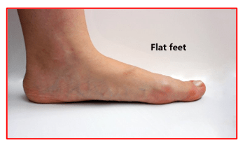
The surgeon then tries to identify if the patient’s flatfoot is flexible or non-flexible (rigid flat feet). This is done by asking the patient to stand on their tiptoes whilst holding onto a table for support.
If the foot arch forms, then the flat foot is called flexible flat foot.
If the arch does not form with standing, it is called rigid flat foot.
The surgeon also examines patient’s both lower leg joints as well the spine for any associated pathologies.
Patients with pain and those with rigid flat feet are advised investigations.
X-rays of the feet in 3 different planes are taken. These may show changes of arthritis, bony fusions (tarsal coalitions) and any deformities of the bones
CT scan also helps look at bones in the foot and can confirm tarsal coalitions missed on x-rays.
MRI scan looks at the tendons in the foot and confirms their health and disease status.
Flat feet treatment involves conservative and surgical methods.
Conservative Treatment:
Flat feet in a child do not need treatment if they are not causing pain or any walking problems.
Children’s feet will grow and develop the same, whether special shoes, shoe inserts, heel cups, or wedges are used. They may walk barefoot, run or jump, or do any other activity without making the flat feet worse.
In older children and adults, flexible flat feet that do not cause pain or walking problems, do not need further treatment.
If they have pain due to flexible flat feet, the following may help:
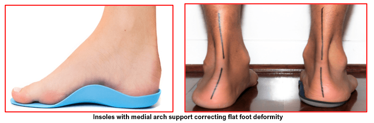
- An arch-support that is used in patient’s shoes. These can be bought at a shoe shop or be custom-made
- Special shoes
- Calf muscle stretches
- Simple painkiller medications as needed
Rigid or painful flat feet needs detailed assessment. The definitive treatment for these depends on the cause of the flat feet.
For tarsal coalition, treatment starts with rest and possibly a cast. Surgery may be needed if pain does not improve.
In more severe cases, surgery may be needed to:
Clean or repair the tendon
Transfer of a tendon to restore the arch
Fuse joints in the foot into a corrected position
Flat feet in older adults can be treated with painkiller medications, insoles with medial arch support, and sometimes surgery.
Flat foot deformity may be surgically treated by correcting any bony deformities and repairing the supporting tendons and ligaments. Surgery is advised for patients not responding to conservative measures and those who have symptomatic tarsal coalitions.
There are various procedures performed under general or spinal anaesthesia.
These include:
- Medializing calcaneal osteotomy:Excision of a portion of the heel bone which is then shifted to the normal anatomic position under the leg and fixed in place with a metal plate and screws.
- Lateral column lengthening:A bone wedge fixed within the outer portion of the heel bone to increase its length and prevent or correct outward rotation of the foot.
- First metatarsal fusion or medial cuneiform dorsal opening wedge osteotomy:An incision is made on the top of the foot and the bones are fused or a portion of bone is removed, and a bony wedge is placed to prevent the big toe portion from raising off the ground.
- Tendon and ligament repair or replacement: This may involve repair or removal of a damaged posterior tibial tendon or lengthening of a tight Achilles tendon. Tendons may be transferred to support the arch. Supporting ligaments are also repaired.
- Double or Triple arthrodesis:Fusion of two or three bones in the hind foot. This is usually performed for advanced stages of the disease characterized by arthritis and stiff deformity.
- Depending on the type of surgery and anaesthesia used, the patients may be allowed to go home the same day or hospitalized for an overnight stay. Operated leg is immobilized in a splint or a cast.
- Leg is kept elevated over 2 pillows to void selling and pain.
- They are taught to mobilise non weight-bearing for 6 to 8 weeks using a pair of elbow crutches or a walker.
- Foot wound is examined, and any sutures removed at 2 weeks.
- A new cast or a removable ankle walker boot may be applied depending on patient’s condition.
- X-rays of the foot and ankle region are taken at 6 weeks and 12 weeks mark to assess correction of deformity and healing of any osteotomy.
As with any surgical procedure, complications can occur and include the risks associated with anaesthesia, infection, swelling, stiffness, damage to blood vessels and nerves, delayed healing, incomplete healing or non-union of bony osteotomies, need for specialised shoes if regular once don’t fit and failure of fixation implants.
These complications are rare and the success rate after flatfoot reconstruction is very high.