Elbow Sprain
Elbow sprain is an injury to the soft tissues of the elbow. It is caused due to stretching or tearing (partial or complete tears) of the ligaments which support the elbow joint. Ligaments are a group of fibrous tissues that connect one bone to another in the body.
Three ligaments are present in the elbow joint: the ulnar collateral ligament, the radial collateral ligament, and the annular ligament. These ligaments provide strength and support to the elbow joint along with the surrounding muscles of the arm and forearm while still allowing motion to occur.
If an injury occurs to the elbow joint, any one of these ligaments may be injured.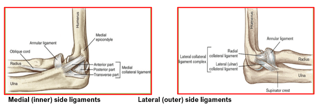
Annular Ligament: courses around the head of the radius bone. It keeps the radius in place when rotating the forearm inwards and outwards.
Ulnar Collateral Ligament (UCL): is a strong fan-shaped condensation of the fibrous joint capsule. It is located on the medial (inner) side of the joint and extends from the medial epicondyle of the humerus to the proximal (upper) portion of the ulna. The UCL protects the elbow against severe valgus stress or pressure from the outside of your arm.
Radial Collateral Ligament: is also a strong fan-shaped condensation of the fibrous joint capsule. It is located on the lateral (outer) side of the joint, extending from the lateral epicondyle of the humerus to the head of the radius. This ligament protects the joint against excessive varus, or inner to outer, stress.
The various causes of an elbow sprain are:
- Involuntary twisting of the arm during sport activities
- Traumatic injury to the elbow due to accidents or a fall
- Overstretching of the elbow during exercise increases tension on the elbow tendons
- Lack of warming up and stretching prior to performing exercises or sports activities
- History of previous elbow sprains makes a patient more vulnerable to another sprain
The common symptoms of elbow sprain include:
- Pain, swelling, tenderness, and bruising around the elbow
- Restricted movement of the elbow
- Pain at the elbow joint while stretching and stressing
The diagnosis of elbow sprain is made based on patient’s history, thorough clinical examination and appropriate investigations.
The elbow will be swollen, bruised, tender and painful to move. There is pain on stressing the joint.
An X-ray of the elbow may be necessary to rule out any fractures or other disease conditions. Rarely, an MRI may be ordered which confirms the elbow sprain and helps in judging the severity of the injury.
Elbow sprains are graded depending upon the severity of the symptoms and clinical examination.
grade I (mild), grade II (moderate) and grade III (severe).
Severe elbow sprains of grade III can lead to elbow dislocation or joint instability.
The treatment for an elbow sprain can be conservative or surgical.
Conservative treatment is as follows:
- Rest: Avoid using the affected elbow for a few weeks. Restrict all activities that cause overuse of the elbow.
- Ice packs: Apply ice bags wrapped in a towel over the sprained elbow for 15-20 minutes at a time to help alleviate any possible pain and swelling.
- Compression: An elastic compression bandage is used to wrap and support the elbow to reduce swelling. Take care not to wrap too tightly which could constrict the blood vessels.
- Elevation: Keep your sprained elbow elevated as much as possible. This can be done by placing pillows under your arm.
- Immobilization: A sling or splint may be applied to stabilize the elbow joint.
- Medications: You will be prescribed pain medications to keep you comfortable, and antibiotics to prevent infection.
- Physiotherapy: Learn appropriate hand exercises that strengthen your forearm muscles. Various modalities of physical therapy such as massage, ultrasound, and muscle stimulation may also be performed to improve muscle strength.
Surgery: Generally, elbow sprains do not require surgery. It is indicated only in cases of severe damage or tear of the ligament.
Elbow sprains and injuries can be prevented by doing the following:
- Exercise on a regular basis to improve muscle strength
- Eat a healthy diet which includes a good variety of nutritious foods
- Use well-checked equipment for any sport activities
- Always warm-up and stretch your muscles prior to performing exercises or sports activities
Ulnar collateral ligament (UCL), also called as the medial collateral ligament, is located on the inside of the elbow and connects the ulna bone to the humerus bone. It is one of the main stabilizing ligaments in the elbow especially with overhead activities such as throwing and pitching.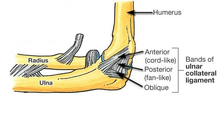
Ulnar collateral ligament (UCL) injury is usually caused by repetitive overhead throwing such as in baseball. The stress of repeated throwing on the elbow causes microscopic tissue tears and inflammation. With continued repetition, eventually the UCL can tear preventing the athlete from throwing with significant speed.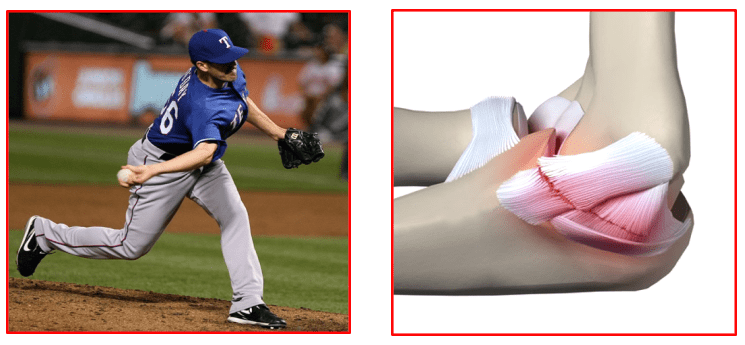
UCL injury may also occur due to direct trauma such as a fall, car accident, or work-related injury. Other causes include any activity that requires repetitive overhead motion of the arm such as tennis, pitching sports, fencing, and painting.
The common symptoms associated with an UCL injury are:
- Pain on inner side of the elbow,
- Unstable elbow joint,
- Numbness in the little finger or ring finger and
- Reduced performance in activities such as throwing baseballs or other objects.
Elbow UCL injury is diagnosed based on patient’s symptoms, thorough clinical examination and appropriate investigations like MRI scan.
The surgeon checks the alignment of the elbow, any tender areas and the range of motion. The UCL is stressed with a valgus or outwards stress to the elbow to reveal the extent of UCL injury and any elbow instability.
Tests like elbow X-rays and MRI scans may be requested to confirm the diagnosis.
UCL injuries can be managed conservatively or surgically.
All patients are generally recommended conservative treatment initially to treat the symptomatic UCL injuries.
Professional athletes, who wish to continue playing sports at a very high level, are offered surgery as first choice of treatment.
Conservative treatment options that are commonly recommended for routine patients include: activity restrictions, orthotics/splints, ice compression, anti-inflammatory medications, analgesic gels, elbow physiotherapy, pulsed ultrasound to increase the blood flow to the injured ligament and promote healing. Patients are also advised to get professional instructions to improve their sports specific techniques and avoid future injuries.
If conservative treatment methods fail to resolve the condition and patient’s symptoms persist beyond 6-12 months, surgery may be recommended for reconstruction of the ulnar collateral ligament.
UCL reconstruction surgery of the elbow repairs the UCL by reconstructing it with a tendon from the patient’s own body (autograft) or from a cadaver (allograft). The most frequently used grafts are the palmaris longus tendon from the patient’s forearm or the hamstring tendons from the knee.
The basic steps for UCL reconstruction surgery include the following:
- The surgery is performed in an operating room under regional or general anaesthesia on a pre-counselled and consented patient.
- An incision is made over the medial epicondyle area
- Care is taken to move muscles, tendons, and nerves out of the way. Extra care is taken to protect the ulnar nerve.
- The donor tendon is harvested from either the forearm or below the knee. The graft is cleaned and prepared
- Drills holes are made into the ulna and humerus bones at pre-determined areas.
- The donor tendon is then inserted through the drilled holes in a figure of 8 pattern
- The tendon is attached to the bone surfaces with special strong sutures
- The incision is closed with absorbable sutures and covered with sterile dressings. Sterile compression bandage is given.
- Finally, a special elbow range of motion splint is applied with the elbow flexed at 90 degrees.
- Patients are sent to the recovery room.
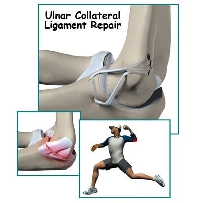
Patients are given appropriate guidelines for post-surgery rehabilitation depending on the type of repair performed and the surgeon’s preference.
Common post-operative guidelines include:
- Elevation of operated arm above heart level to reduce swelling and pain
- Wear an immobilizing splint for 1-3 weeks
- Apply ice packs to the surgical area 4-5 times to reduce swelling and pain
- Keep the surgical wound clean and dry. Cover the area with plastic wrap when bathing or showering.
- Physiotherapy is started early for strengthening and stretching exercises which help in regaining elbow mobility, improving muscle strength and giving better elbow function. Professional athletes can expect a strenuous strengthening and range of motion rehabilitation programme for 6-12 months before returning to their sport
Eating a healthy diet and not smoking will promote healing
The majority of patients suffer from no complications following UCL reconstruction surgery. However, complications can occur following elbow surgery and include swelling, infection, stiffness or limited range of motion, nerve damage causing numbness, tingling, burning or loss of sensations in the hand and forearm area, cubital tunnel syndrome and elbow instability.
Book An Appointment
Private Clinics : Locations & Directions
London Joints Clinic (Pune)
Address
Office S 5, 2nd Floor, North Block, Sacred World Mall,
Opp Sacred Heart Township, Near Jagtap Chowk,
Wanawadi, Pune 411040
Monday to Saturday
6 PM to 9 PM
Appointments
Hospitals OPDs : Locations & Directions
Jupiter Hospital (Baner)

Address
Lane 3, Baner- Balewadi Road,
Prathamesh Park,
Baner, Pune 411 045
Monday to Saturday 11 AM to 4 PM
Appointments
Contact us
Dr Anand Jadhav has a centralised appointment system for all locations across various hospitals and clinics in Pune & PCMC areas
Appointment Bookings & Requests can be made by any method :

